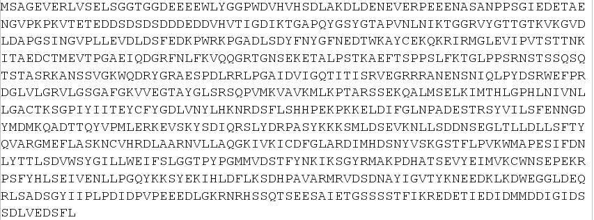
In the image above you can see the sequence of the FIP1L1-PDGFRA fusion protein. If you pass the mouse over the picture you will see the exons which come from FIP1L1 in dark green and blue, and the exons which come from the PDGFRA in soft green and yellow. In red you will see the last part of the exon number 12 of PDGFRA. You can compare this image with the ones you can see in the other links to see how the protein has been made.