RPS-BLAST 2.2.5 [Nov-16-2002]
Query= local sequence:
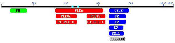
hg13_dna|geneid_v1.1_predicted_protein_2|1064_AA
(1063 letters)
Database: cdd.v1.60
10,013 PSSMs; 2,494,783 total columns
 Ā
Ā ĀĀ .. This CD alignment includes 3D structure. To display structure, download
Cn3D! ĀĀ .. This CD alignment includes 3D structure. To display structure, download
Cn3D!
| Ā |
PSSMs producing significant alignments: |
Score
(bits) |
E
value |
| Ā |
|
gnl|CDD|5336 |
cd00137, PLCc, Phospholipase C, catalytic domain; Phosphoinosi... |
284 |
4e-77 |
|
gnl|CDD|133 |
smart00148, PLCXc, Phospholipase C, catalytic domain (part); d... |
174 |
4e-44 |
|
gnl|CDD|134 |
smart00149, PLCYc, Phospholipase C, catalytic domain (part); d... |
167 |
5e-42 |
|
gnl|CDD|951 |
pfam00388, PI-PLC-X, Phosphatidylinositol-specific phospholipa... |
144 |
5e-35 |
|
gnl|CDD|950 |
pfam00387, PI-PLC-Y, Phosphatidylinositol-specific phospholipa... |
139 |
2e-33 |
|
gnl|CDD|14861 |
cd00275, C2_2, Protein kinase C conserved region 2, subgroup 2... |
120 |
6e-28 |
|
gnl|CDD|8952 |
smart00239, C2, Protein kinase C conserved region 2 (CalB); Ca... |
78.3 |
4e-15 |
|
gnl|CDD|14771 |
cd00030, C2, Protein kinase C conserved region 2 (CalB); Ca2+-... |
75.1 |
3e-14 |
|
gnl|CDD|9086 |
pfam00168, C2, C2 domain |
71.9 |
3e-13 |
|
gnl|CDD|14862 |
cd00276, C2_1, Protein kinase C conserved region 2, subgroup 1... |
51.0 |
7e-07 |
| Ā |
gnl|CDD|14168 |
COG5038, COG5038, COG5038, Ca2+-dependent lipid-binding protei... |
40.7 |
8e-04 |
|
gnl|CDD|9087 |
pfam00169, PH, PH domain. PH stands for pleckstrin homology |
37.5 |
0.007 |
|
gnl|CDD|5336,
cd00137, PLCc, Phospholipase C, catalytic domain; Phosphoinositide-specific
phospholipases C catalyze hydrolysis of phosphatidylinositol-4,5-bisphosphate
(PIP2) to D-myo-inositol-1,4,5-trisphosphate (1,4,5-IP3) and sn-1,2-diacylglycerol
(DAG). Both products function as second messengers in eukaryotic signal transduction
cascades. 1,4,5-IP3 triggers inflow of calcium from intracellular stores;
the membrane resident product DAG controls cellular protein phosphorylation
states by activating various protein kinase C isozymes. The enzyme comprises
2 regions (X and Y) connected via a linker which may contain inserted domains,
X and Y together form a TIM barrel-like structure containing the active site
residues. |
             CD-Length = 298 residues, 100.0% aligned
             Score =  284 bits (727), Expect = 4e-77
Query:  331   KVCQDMKQPLSHYFINSSHNTYLIEDQFRGPSDITGYIRALKMGCRSVELDVWDGPDNEP  390
Sbjct:  1     MVAQDMSKPLSHYFIPSSHNTYLTGKQVWGESSIEGYIQALKHGCRCVELDCWDGPDNEP  60

Query:  391   VIYTGHTMTSQIVFRSVIDIINKYAFFASEYPLILCLENHCSIKQQKVMVQHMKKLLGDK  450
Sbjct:  61    VVYHGHTFTTPIKLSEVLEAIKDFAFVTSPYPVILSLEDHCSPDQQAKMADSFKETFGDL  120

Query:  451   LYTTSPNVEESYLPSPDVLKGKILIKAKKLSSNCSGVEGDVTDEDEGAEMSQRMGKENME  510
Sbjct:  121   LYTPPTFSSLNVLPSPE-----QLKGKILLKGKKSGTYLDALEKEEGDSSQHSDSSESMS  175

Query:  511   QPNNVPVKRFQLCKELSELVSICKSVQFKEFQ-VSFQVQKYWEVCSFNE--VLASKYANE  567
Sbjct:  176   SEKKPSEKTHIRIAPESSELIGYQSLQWKDGETFTTESNQSLNIFSQSEYKVLLVKAIKE  235

Query:  568   NPGDFVNYNKRFLARVFPSPMRIDSSNMNPQDFWKCGCQIVAMNFQTPGLMMDLNIGWFR  627
Sbjct:  236   TPLKLVKTNQNYLLRVYPSGTRGDSSNYNPQIAWNAGVQIVALNFQTYGEGMQLNLGMFR  295

Query:  628   QNG  630
Sbjct:  296   ANG  298
|
gnl|CDD|133,
smart00148, PLCXc, Phospholipase C, catalytic domain (part); domain X; Phosphoinositide-specific
phospholipases C. These enzymes contain 2 regions (X and Y) which together
form a TIM barrel-like structure containing the active site residues. Phospholipase
C enzymes (PI-PLC) act as signal transducers that generate two second messengers,
inositol-1,4,5-trisphosphate and diacylglycerol. The bacterial enzyme appears
to be a homologue of the mammalian PLCs. |
             CD-Length = 145 residues,  92.4% aligned
             Score =  174 bits (443), Expect = 4e-44
Query:  334   QDMKQPLSHYFINSSHNTYLIEDQFRGPSDITGYIRALKMGCRSVELDVWDGPDNEPVIY  393
Sbjct:  1     QDMSKPLSHYFINSSHNTYLTGKQLWGESSVEGYIQALKHGCRCVELDCWDGPDGEPVIY  60

Query:  394   TGHTMTSQIVFRSVIDIINKYAFFASEYPLILCLENHCSIKQQKVMVQHMKKLLGDKLYT  453
Sbjct:  61    HGHTFTLPIKLSEVLEAIKKFAFVTSPYPVILSLENHCSPDQQAKMAQMFKEIFGDLLYT  120

Query:  454   TSPNVEESYLPSPD  467
Sbjct:  121   PPTTSSLEYLPSPE  134
|
gnl|CDD|134,
smart00149, PLCYc, Phospholipase C, catalytic domain (part); domain Y; Phosphoinositide-specific
phospholipases C. These enzymes contain 2 regions (X and Y) which together
form a TIM barrel-like structure containing the active site residues. Phospholipase
C enzymes (PI-PLC) act as signal transducers that generate two second messengers,
inositol-1,4,5-trisphosphate and diacylglycerol. The bacterial enzyme appears
to be a homologue of the mammalian PLCs. |
             CD-Length = 117 residues, 100.0% aligned
             Score =  167 bits (425), Expect = 5e-42
Query:  526   LSELVSICKSVQFKEFQVSFQVQKYWEVCSFNEVLASKYANENPGDFVNYNKRFLARVFP  585
Sbjct:  1     LSELVSYCAPVKFRSFELAEEKNPFYEMSSFSETKAKKLLEKAPTDFVRYNQRQLSRVYP  60

Query:  586   SPMRIDSSNMNPQDFWKCGCQIVAMNFQTPGLMMDLNIGWFRQNGNCGYVLRPAIMR  642
Sbjct:  61    KGTRVDSSNYNPQVFWNHGCQMVALNFQTPDKAMQLNQGMFRANGGCGYVLKPDFLR  117
|
gnl|CDD|951,
pfam00388, PI-PLC-X, Phosphatidylinositol-specific phospholipase C, X domain.
This associates with pfam00387 to form a single structural unit. |
             CD-Length = 145 residues,  91.7% aligned
             Score =  144 bits (365), Expect = 5e-35
Query:  335   DMKQPLSHYFINSSHNTYLIEDQFRGPSDITGYIRALKMGCRSVELDVWDGPDNEPVIYT  394
Sbjct:  1     DMSIPLSHYFISSSHNTYLTGKQLWGKSQVESYRQQLDHGCRCVELDCWDGPDDEPIIYH  60

Query:  395   GHTMTSQIVFRSVIDIINKYAFFASEYPLILCLENHCSIKQQKVMVQHMKKLLGDKLYTT  454
Sbjct:  61    GGTFTLEIKLKDVLEAIKDFLFKTSPYPIILSLENHCNSDQQRKMAKYFEEIFGDYLLTK  120

Query:  455   SPNVEESY-LPSPD  467
Sbjct:  121   -PLDSLTTKLPSLK  133
|
gnl|CDD|950,
pfam00387, PI-PLC-Y, Phosphatidylinositol-specific phospholipase C, Y domain.
This associates with pfam00388 to form a single structural unit. |
             CD-Length = 118 residues, 100.0% aligned
             Score =  139 bits (351), Expect = 2e-33
Query:  525   ELSELVSICKSVQFKEFQVSFQVQKYWEVCSFNEVLASKYANENPGDFVNYNKRFLARVF  584
Sbjct:  1     ELSNLVNYIQSIKFRSFSLPTEKNTSYEMSSFSERKAKQLLKESPIEFVKHNKRQLSRVY  60

Query:  585   PSPMRIDSSNMNPQDFWKCGCQIVAMNFQTPGLMMDLNIGWFRQNGNCGYVLRPAIMR  642
Sbjct:  61    PKGTRFDSSNFMPQPFWNAGCQMVALNFQTSDLPMQINLGMFEYNGGSGYLLKPPFLR  118
|
gnl|CDD|14861,
cd00275, C2_2, Protein kinase C conserved region 2, subgroup 2; C2 Ca2+-binding
motif present in phospholipases, protein kinases C, and synaptotagmins (amongst
others); some PKCs lack calcium dependence. Particular C2s appear to bind
phospholipids, inositol polyphosphates,and intracellular proteins. Two distinct
C2 topologies generated by permutation of the sequence with respect to the
N- and C-terminal beta strands are seen. In this subgroup, containing phospholipases
C and D( PLC-1, PLD) and specific protein kinases C (PKC) subtypes, the C-terminal
beta strand occupies the position of what is the N-terminal strand in subgroup
1. |
             CD-Length = 129 residues,  99.2% aligned
             Score =  120 bits (303), Expect = 6e-28
Query:  664   LHIKIISGQNFPKPKGS--GAKGDVVDPYVYVEIHGIPADCAEQRTKTVHQNGDAPIFDE  721
Sbjct:  2     LKVRIIEAQQLPKTDKSLDGKSKSTLDPYVTVEIDGVDA--RIGRTKVIQNNGFNPVWNE  59

Query:  722   SFEFQINLPELAMVRFVVLDDDYI-GDEFIGQYTIPF-ECLQTGYRHVPLQSLTGEVLAH  779
Sbjct:  60    EFEFPLAHPDLAFIRFTVKDSDPIGGDDFIGNATIPVDEVGKQGYRWIPLLDMNGEQLPF  119

Query:  780   ASLFVHVAIT  789
Sbjct:  120   SKLFVKIQLE  129
|
gnl|CDD|8952,
smart00239, C2, Protein kinase C conserved region 2 (CalB); Ca2+-binding
motif present in phospholipases, protein kinases C, and synaptotamins (among
others). Some do not appear to contain Ca2+-binding sites. Particular C2s
appear to bind phospholipids, inositol polyphosphates, and intracellular
proteins. Unusual occurrence in perforin. Synaptotagmin and PLC C2s are permuted
in sequence with respect to N- and C-terminal beta strands. SMART detects
C2 domains using one or both of two profiles. |
             CD-Length = 101 residues, 100.0% aligned
             Score = 78.3 bits (192), Expect = 4e-15
Query:  663   LLHIKIISGQNFPKPKGSGAKGDVVDPYVYVEIHGIPADCAEQRTKTVHQNGDAPIFDES  722
Sbjct:  1     TLTVKIISARNLPPKDKGGK----SDPYVKVSLDGDPREKK--KTKVVKNTLN-PVWNET  53

Query:  723   FEFQINLPELAMVRFVVLDDDYIG-DEFIGQYTIPFECLQTGYRHVPL  769
Sbjct:  54    FEFEVPPPELSELEIEVYDKDRFSRDDFIGQVTIPLSDLLLGGRHEKL  101
|
gnl|CDD|14771,
cd00030, C2, Protein kinase C conserved region 2 (CalB); Ca2+-binding motif
present in phospholipases, protein kinases C, and synaptotagmins (among others).
Some do not appear to contain Ca2+-binding sites. Particular C2s appear to
bind phospholipids, inositol polyphosphates,and intracellular proteins. Synaptotagmin
and PLC C2s are permuted in sequence with respect to N- and C-terminal beta
strands. |
             CD-Length = 105 residues, 100.0% aligned
             Score = 75.1 bits (184), Expect = 3e-14
Query:  663   LLHIKIISGQNFPKPKGSGAKGDVVDPYVYVEIHGIPADCAEQRTKTVHQNGDAPIFDES  722
Sbjct:  1     RLTVKIIEARNLPPKDKKGT----SDPYVKVSLGGDKK--QKKKTKVV-KKTLNPVWNET  53

Query:  723   FEFQINLPELAMVRFVVLDDDYIG-DEFIGQYTIPFECL----QTGYRHVPL  769
Sbjct:  54    FTFEVPPPEESSLVIEVYDYDKFSRDDFIGEVTIPLSELLLDGKEGDRWFPL  105
             CD-Length = 85 residues, 100.0% aligned
             Score = 71.9 bits (176), Expect = 3e-13
Query:  664   LHIKIISGQNFPKPKGSGAKGDVVDPYVYVEIHGIPADCAEQRTKTVHQNGDAPIFDESF  723
Sbjct:  1     LRVTVIEAKNLPPKDKNGK----SDPYVKVSLGGQKKDT--KKTKVIKNTLN-PVWNETF  53

Query:  724   EFQINLPELAMVRFVVLDDDYIG-DEFIGQYT  754
Sbjct:  54    TFEVPPPELAELRIEVYDYDRFGKDDFIGEVS  85
|
gnl|CDD|14862,
cd00276, C2_1, Protein kinase C conserved region 2, subgroup 1; C2 Ca2+-binding
motif present in phospholipases, protein kinases C, and synaptotagmins (amongst
others); some PKCs lack calcium dependence. Particular C2s appear to bind
phospholipids, inositol polyphosphates,and intracellular proteins. Two distinct
C2 topologies generated by permutation of the sequence with respect to the
N- and C-terminal beta strands are seen. In this subgroup, containing synaptotagmins,
specific protein kinases C (PKC) subtypes and other proteins, the N-terminal
beta strand occupies the position of what is the C-terminal strand in subgroup
2. |
             CD-Length = 124 residues,  75.0% aligned
             Score = 51.0 bits (122), Expect = 7e-07
Query:  662   QLLHIKIISGQNFPKPKGSGAKGDVVDPYVYVEIHGIPADCAEQRTKTVHQNGDAPIFDE  721
Sbjct:  15    GQLTVVIIKARNLPPMDKNGL----SDPYVKVYLLPDGKKKKKKKTK-VKRKTLNPVFNE  69

Query:  722   SFEFQINLPELAMVR--FVVLDDDYIG-DEFIGQYTIP  756
Sbjct:  70    TFVFDVPPEELADRSLQITVWDYDRFSRNDFIGEVVIG  107
gnl|CDD|14168, COG5038, COG5038, Ca2+-dependent lipid-binding protein, contains C2 domain [General function prediction only]
             CD-Length = 1227 residues, only   7.5% aligned
             Score = 40.7 bits (95), Expect = 8e-04
Query:  664   LHIKIISGQNFPKPKGSGAKGDVVDPYVYVEIHGIPADCAEQRTKTVHQNGDAPIFDESF  723
Sbjct:  1042  LTIMLRSGENLP----SSDENGYSDPFVKLFLNE-----KSVYKTKVVKKTLNPVWNEEF  1092

Query:  724   EFQINLPELAMVRFVVLDDDYIG-DEFIGQYTIPFECLQTG  763
Sbjct:  1093  TIEVLNRVKDVLTINVNDWDSGEKNDLLGTAEIDLSKLEPG  1133
|
gnl|CDD|9087, pfam00169, PH, PH domain. PH stands for pleckstrin homology.
|
             CD-Length = 100 residues,  97.0% aligned
             Score = 37.5 bits (86), Expect = 0.007
Query:  53    EGSELKKVRSNSRIYH-RYFLLDADMQSLRWEPSKKDSEKAKIDIKSIKEVRTGKNTDIF  111
Sbjct:  4     EGWLLKKSTVKKKRWKKRYFFLFNDVLIYYKDKKKSYEPKGSIPLSGC-SVEDVPDSEF-  61

Query:  112   RSNGISDQISEDCAFSVIYGENYESLDLVANSADVANIWVTGLRYLIS  159
Sbjct:  62    ---------KRPNCFQLRSRDGKETFILQAESEEERQDWIKAIQSAIR  100
|